Whether you are making a visit to the doctor’s office, ER, or chiropractor, there are times when you will need to have medical imaging done. This imaging allows professionals to see what is going on inside your body. Imaging was revolutionary for the medical world and chiropractic world alike. Imaging allows for your care provider to get a solid idea on what may be causing you issues, and how they can help solve these problems. Chiropractors use imaging mainly to see the structure of your bones, as well as to see if they can spot any subluxations. There are multiple types of imaging you may be ordered to get and knowing what each type of imaging does can help you understand the process your chiropractor is leading you through a bit better.
X-ray
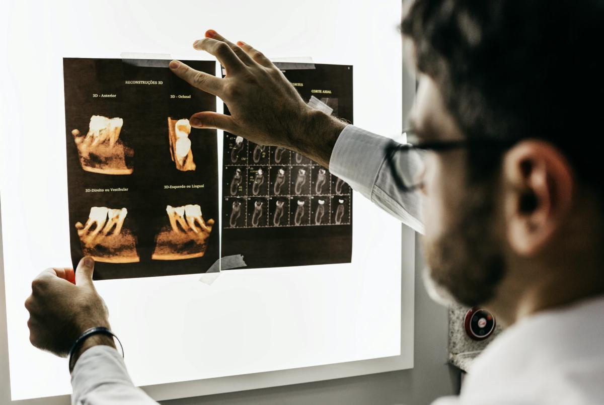
An X-Ray is probably the type of imaging you are most familiar with. An X-ray involves radioactive waves passing through the body that only capture hard tissue, such as bones. The X-ray machine then transfers the result to an image that can be studied and understood. X-rays are commonly used by dentists and orthodontists for your teeth, as well as to diagnose broken bones and bone tumors. This is also the form of imaging most used by chiropractors as it gives them the information they need on your bone structure to come up with the correct treatment plan.
Computerized Tomography (CT)
A CT scan is a more sensitive version of an x-ray. It uses additional software to create 3D images of soft tissue surrounding the bones. CT scans are typically used by radiologists to locate soft tissue tumors as well as detect their size. They are also used to detect different types of cancer that an x-ray would not pick up. It is also just as short as an x-ray, only lasting roughly 10-15 minutes.
Magnetic Resonance Imaging (MRI)
MRIs are different from x-rays and CT scans in that they use different waves to achieve the image they are looking for. An MRI uses a mixture of magnetic fields and radio waves to create a detailed image of the organs and soft tissue. This is the type of imaging you always see in medical shows, where the patient is laying down and slide into a large tube that is emitting a magnetic field. This is also a commonly used type of imaging by chiropractors. It can be used to detect things in the brain such as an aneurysm, as well as things such as a stroke, MS, or tumors. Chiropractors mainly use it to diagnose spinal disorders, as it creates a vivid image of the shape and state of the spinal cord.
Positron Emission Tomography (PET)
A PET scan is when radioactive tracers are used to detect irregularities in body tissue. It is often used in combination with CT or MRI scans. PET scans are used to monitor the progress of different conditions such as cancer, diagnose degenerative diseases, and plan surgeries.
Ultrasound
Ultrasounds involve the use of high frequency sound waves to create images of soft tissue, muscle, and organs. This type of imaging is most often used to check on the progress of an unborn baby and to plan surgeries.
Using the right kind of medical imaging can give your doctor the information they need to most effectively diagnose and treat your condition. Duncan Chiropractic starts all new patients with X-Rays to determine the current state of your skeletal system, but sometimes other types of imaging are required for more in-depth diagnoses. At this point, we will refer you out for that imaging to create the best treatment plan for your situation.